Who is not familiar with dogs? They come in many varieties - from beloved characters like Maxipes Fík, Štaflík, and Špagetka, to historical figures like Lajka, and form stray dogs to purebred dogs with pedigrees. The International Canine Federation registers over 340 breeds, representing a diverse mix of sizes, colours, and qualities. As human companions, dogs have earned their own international day, which has been celebrated annually on 26 August since 2004.
Dogs were domesticated more than 10,000 years ago. Since then, they have accompanied and protected their masters, guarded herds, assisted persons with impaired vision, checked for explosives and drugs, and helped track down criminals... However, most importantly, they are living creatures that can sometimes suffer from diseases.
One group of these diseases are tumours, including mastocytomas —mast cell tumours. Mastocytomas are among the most common canine tumours, accounting for up to 21% of all canine skin tumours. Their appearance, manifestations, and development are varied, ranging from stable, slow-growing forms to aggressive, metastatic ones, making prognosis difficult to determine. A significant negative prognostic factor is the presence of mutations in the c-KIT gene. However, detection of the mutation of c-KIT gene is a direct indication for therapy with targeted drugs that minimize the negative effects of conventional chemotherapy, improve quality of life and prolong survival even in dogs with inoperable mastocytomas.
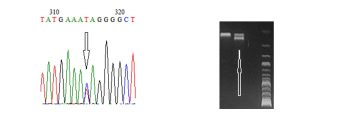
In laboratories of the Veterinary Research Institute, we perform detection of c-KIT mutations by PCR followed by sequencing. We offer veterinarians in clinical practice c-KIT gene assessment in canine mastocytoma samples, identifying either point mutations (Figure 1) or duplications (Figure 2).

Figure 1: Detection of point mutation in the c-KIT gene by sequencing Figure 2: Detection of duplication in the c-KIT gene on gel
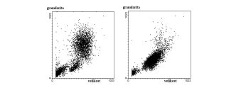
The other group of tumours includes haematological malignancies, particularly leukaemias and lymphomas. These are characterized by uncontrolled proliferation of cells, primarily lymphocytes, in the bone marrow or lymphoid tissue, such as lymph nodes. Owing to advances in small animal veterinary medicine, these diseases are now treatable even in these patients. Since there are several types of lymphocytes that differ in their functions, it is crucial to identify the specific cell type from which these tumours originate in order to tailor therapy. One of the basic laboratory techniques used for the diagnosis is flow cytometry. Flow cytometry enables the detection of the morphological characteristics of cells in a sample, allowing for the identification of pathological populations (Fig. 3). Using monoclonal antibodies against various CD markers facilitates the determination of whether the pathological cells are developmentally younger — cells that generally respond better to therapy — or developmentally older, cells that proliferate more slowly but are more resistant to treatment. The laboratories at the Veterinary Research Institute analyse over 120 such samples annually, aiding veterinarians in making more informed decisions for their patients.

Figure 3: Morphological characteristics of cells in the blood of a healthy dog (left) and a dog affected by acute lymphoblastic leukaemia (right), as analysed by flow cytometry.


